FIGURE 1.

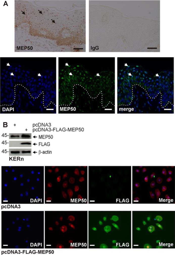
MEP50 is expressed in the human epidermis and in cultured keratinocytes. A, foreskin tissue sections were stained with anti-MEP50, and binding was visualized with peroxidase-conjugated secondary antibody (top panels). The control is IgG. The arrows indicate MEP50 nuclear localization in suprabasal keratinocytes. Foreskin tissue sections (bottom panels) were fixed and stained with anti-MEP50, and antibody binding was visualized using a FITC-conjugated secondary antibody. The arrows indicate nuclear MEP50 accumulation. Scale bars = 10 μm. B, colocalization of endogenous and expressed MEP50. KERn were electroporated with 3 μg of pcDNA3 or pcDNA3-FLAG-MEP50. After 48 h, protein lysates were tested by immunoblot using anti-FLAG and anti-MEP50. β-actin was used as the loading control. After 48 h, the cells were fixed and costained with anti-FLAG (green) and anti-MEP50 (red). Similar results were observed in each of three experiments. The staining indicates MEP50 distribution in the nucleus and cytoplasm. Scale bars = 10 μm.
