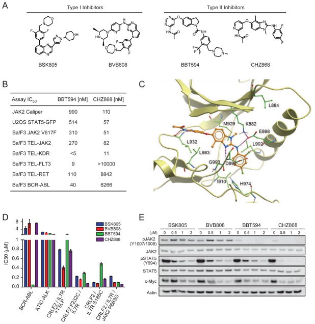
Figure 1. The type II JAK2 inhibitor NVP-CHZ868 blocks JAK2 signaling in vitro and in vivo.
(A) Chemical structures of type I and II JAK2 inhibitors. (B) IC50 values for CHZ868 and BBT594 in enzymatic and cell-based assays. (C) Binding mode model of CHZ868 to JAK2. Ribbon representation of the JAK2 kinase domain with CHZ868 illustrated as a stick model. Amino acid side chains interacting with the inhibitor are shown in green. Polar contacts between the protein and the inhibitor are highlighted with dotted purple lines. (D) IC50 values for type I and II JAK2 inhibitors in Ba/F3 cells expressing the indicated proteins in the absence of cytokines, except where “+TSLP” indicates 1 nM TSLP. Error bars represent SEM. (E) Immunoblotting against the indicated targets using lysates from Ba/F3-CRLF2/JAK2 R683G cells exposed to the indicated concentrations of JAK2 inhibitors for 2 hr. See also Figure S1 and Table S1.

