FIG 5.
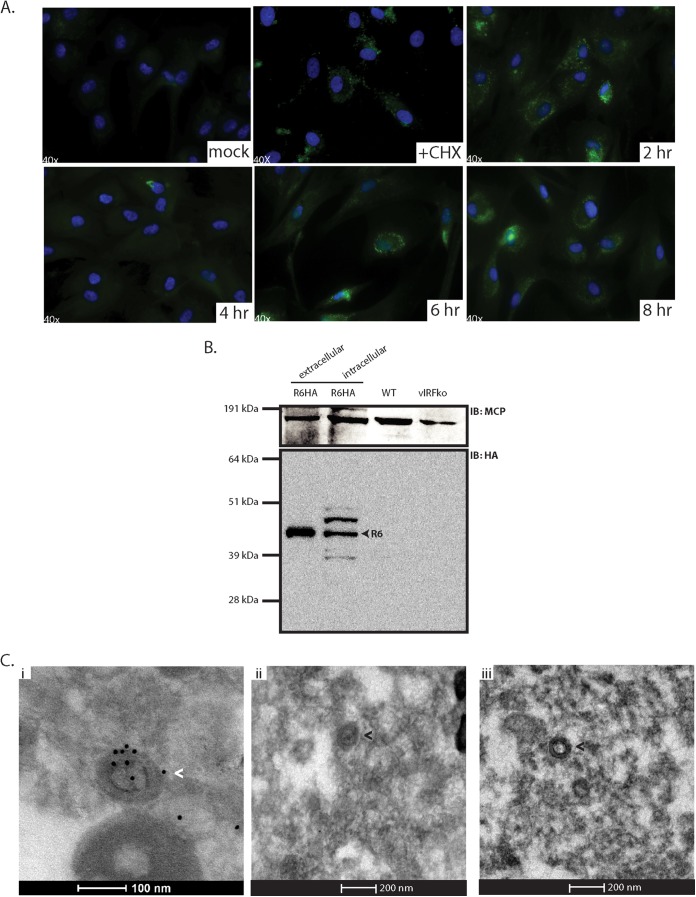
R6 is associated with RRV virions. (A) Primary RFs were pretreated with CHX and subsequently infected with R6HA-RRV at an MOI of 2.5. CHX was removed, and the cells were fixed at the indicated time points and analyzed by immunofluorescence for the detection of R6-HA (anti-HA) (green) and stained with Hoechst (blue) for the detection of nuclei. (B) 1 × 10⁵ PFU of gradient-purified virus samples (extracellular R6HA-RRV, intracellular R6HA-RRV, WTBACRRV, and vIRFko-RRV) was subjected to SDS-PAGE and probed with anti-HA antibody and anti-MCP antibody as a control. (C) (i) Gradient-purified R6HA-RRV was fixed, pelleted, and sectioned. The sections were immunogold stained with anti-HA antibody and 10-nm gold-conjugated secondary antibody. (ii) Gradient-purified WTBACRRV was sectioned and immunogold stained with anti-HA antibody and 10-nm gold-conjugated secondary antibody as a control. (iii) R6HA-RRV sections were stained with 10-nm gold-conjugated secondary antibody alone as a control. Virus particles with gold particles are indicated by the white arrowhead, and virus particles with no gold particles are indicated by black arrowheads.

