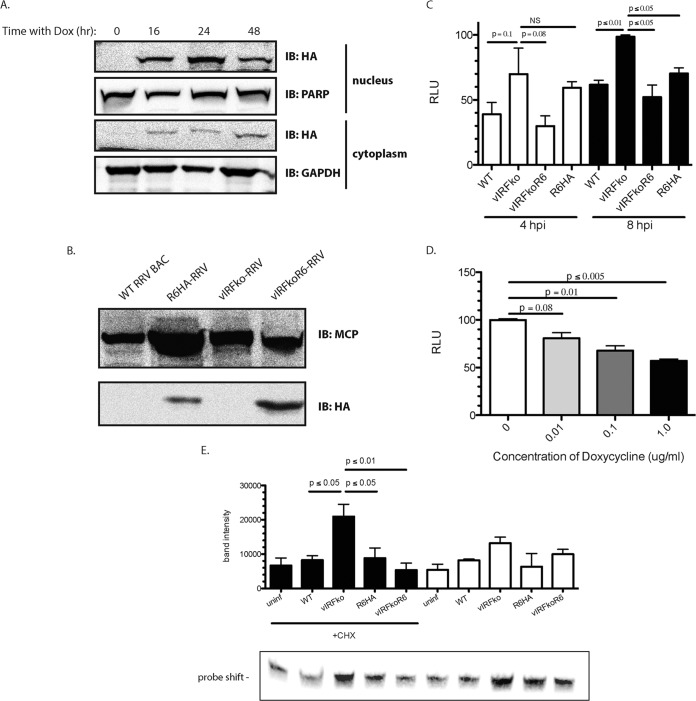
FIG 6.
Virion-associated R6 is functional. (A) Telomerized RF-rtTAs stably transduced with R6-HA were treated with Dox for the indicated times. The nuclear and cytoplasmic lysates were subjected to SDS-PAGE and probed with anti-HA antibody. GAPDH and PARP served as loading controls and as controls for purity of fractionation for the cytoplasmic and nuclear extracts, respectively. (B) Telomerized RF-rtTAs were treated with Dox and infected with vIRFko-RRV at an MOI of 0.01 per cell. The resultant virus (vIRFkoR6-RRV) was gradient purified from cell supernatants. Five micrograms of gradient-purified virus (WTBACRRV, vIRFko-RRV, R6HA-RRV, and vIRFkoR6-RRV) was subjected to SDS-PAGE and probed with anti-HA antibody and anti-MCP antibody as a control. (C) tRF-ISREs were infected for 4 or 8 h with the indicated virus at an MOI of 2.5 PFU per cell. The cells were then assayed for firefly luciferase activity. Firefly luciferase levels were normalized to constitutively expressed Renilla luciferase levels in each well. The data are averages (and SEM) from the results of 3 independent experiments. (D) tRF-rtTA:R6HAs were infected with vIRFko-RRV at an MOI of 0.01 PFU per cell after pretreatment with the indicated amounts of Dox. Virus was then harvested and gradient purified. The resultant virus was used to infect tRF-ISREs at an MOI of 2.5 PFU per cell for 8 h. The cells were assayed for firefly luciferase activity. Firefly luciferase levels were normalized to constitutively expressed Renilla luciferase levels in each well. The data are averages (and SEM) from the results of 3 independent experiments. The total levels of MCP and R6HA in virion preparations were assessed by Western blotting with anti-MCP and anti-HA antibodies. (E) Telomerized RFs were infected with the indicated virus at an MOI of 2.5 PFU per cell for 8 h in the presence or absence of CHX. EMSA was performed on nuclear extracts (20 μg). The biotin-labeled probe corresponded to the PRDI-PRDIII motif (5′-GAAAACTGAAAGGAGAACTGAAAGTG-3′) of the IFN-β promoter. The data were analyzed using a paired t test. P values of ≤0.05 were considered significant, and values greater than 0.05 were not significant.

