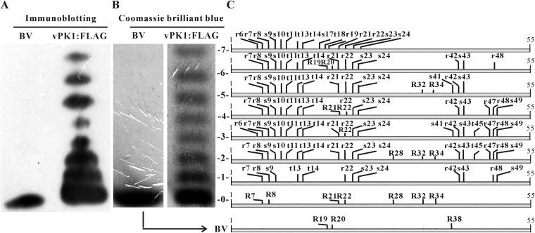
FIG 1.
PTM sites of P6.9. (A) Immunoblotting for P6.9 in vPK1:FLAG-infected cells. vPK1:FLAG-infected cells were harvested at 48 h p.i. P6.9 species were separated by AU-PAGE into 8 rungs based on their charge states and detected by immunoblotting with the anti-P6.9 antibody. (B) The P6.9 species were stained with Coomassie brilliant blue and numbered 0 for the unphosphorylated species (including the BV) and 1 to 7 for the phosphorylated species. (C) All of the identified phosphorylation and methylation sites of P6.9 are indicated in a combined schematic. The phosphorylated residues are indicated by lowercase letters; the methylated residues are indicated by capital letters. vPK1:FLAG, vPK1:FLAG-infected cells; BV, BVs purified from the supernatants of vPK1:FLAG-infected cells; S, serine; T, threonine; R, arginine.

