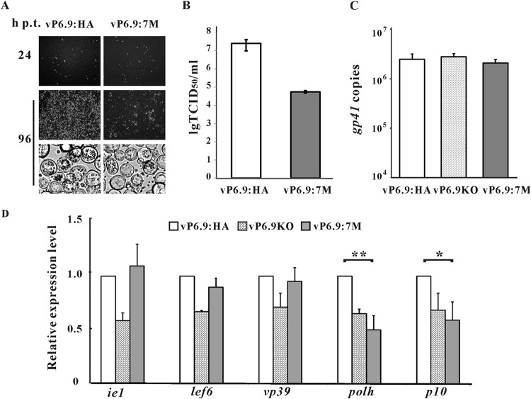
FIG 5.
Hyperphosphorylation of P6.9 regulates viral proliferation and transcription of very late genes. (A) Sf9 cells were transfected with 2 μg bacmid DNA of vP6.9:7M or vP6.9:HA. At 24 and 96 h p.t., cells were observed with a fluorescence microscope or a light microscope. (B) Infectious BV production of vP6.9:7M and vP6.9:HA. The supernatants at 96 h p.t. were harvested, and the BV titers were determined with TCID50 assays. Each value represents the averages from three independent transfections. lg TCID50/ml, the logarithm of the TCID50/ml value with base 10. (C) qPCR analysis of viral DNA replication. Sf9 cells (1 × 10⁶) were transfected with 1 μg bacmid DNA of vP6.9:7M, vP6.9KO, or vP6.9:HA. At 24 h p.t., total cellular DNA was extracted, digested with DpnI, and analyzed by qPCR. Error bars indicate standard deviations for three independent experiments. (D) qRT-PCR analysis of viral gene transcription. Sf9 cells (1 × 10⁶) were transfected with 1 μg bacmid DNA of vP6.9:7M, vP6.9KO, or vP6.9:HA. At 24 h p.t., the indicated viral genes were measured by qRT-PCR. The transcription levels were normalized to that of the host 18S rRNA transcripts and are shown as the percentages of the corresponding genes in the vP6.9:HA bacmid-transfected cells. Data were analyzed by Student's t test. *, P < 0.05; **, P < 0.01. Error bars indicate standard deviations from three independent experiments.

