
FIG 1.
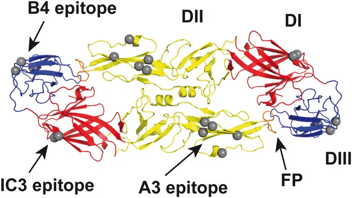
Ribbon diagram of the soluble E protein dimer of TBE virus (Protein Data Bank [PDB] code 1SVB; top view) with the location of epitopes of MAbs used in this study. Structural domains of sE are colored in red (DI), yellow (DII), and blue (DIII) and the fusion peptide (FP) in orange. Gray spheres indicate Cα atoms of residues involved in the binding of MAbs IC3, A3, and B4, as determined by the use of virus escape mutants (76, 77) and engineered recombinant E mutants (52).