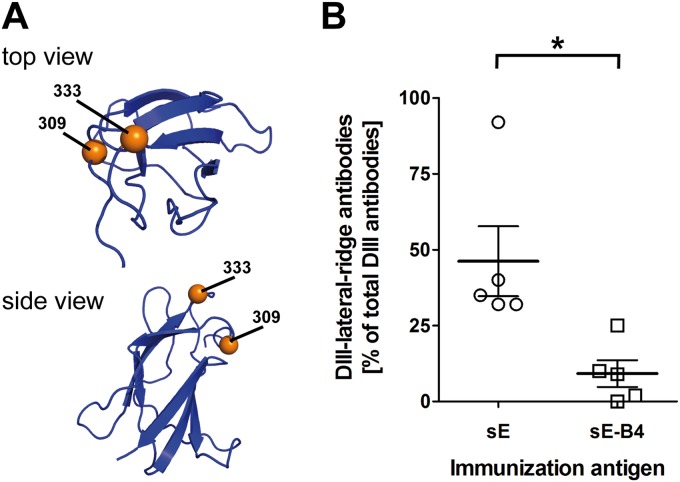
FIG 5.
(A) Ribbon diagram of DIII (PDB code 1SVB) in top and side views. The DIII mutant contains amino acid substitutions at positions 309 and 333, indicated as orange spheres. (B) Antibody response to the DIII-lateral-ridge epitope. Percentages of DIII-lateral-ridge ELISA antibodies in sera of mice immunized with sE or the IC sE-B4. The black star indicates a statistically significant difference between the values for the two groups (unpaired t test; *, P < 0.05).

