
FIG 9.
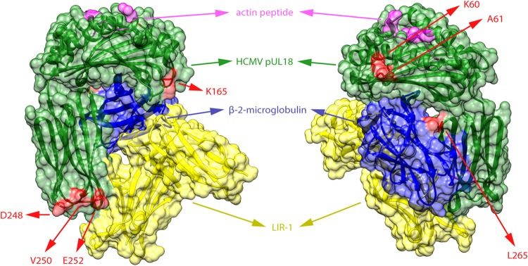
Residues under diversifying selection in pUL18. Codons under positive/diversifying selection in the UL18 gene were determined with the SLAC, FEL, MEME, and FUBAR algorithms of the HyPhy package. Sites that showed significant evidence of positive selection by at least two of four methods are represented in red on the protein structure of pUL18 (green). The structure shows a complex of pUL18 (a viral MHC-I homolog), human β-2-microglobulin (blue) (a MHC-I light chain), and an actin peptide (pink) bound to the inhibitory immunoglobulin receptor LIR-1 (yellow) (136). The three-dimensional structure is visualized from two opposite angles. All selected residues are located at the surface of pUL18.