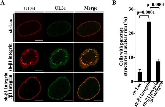
FIG 10.

Effect of β1 integrin on localization of UL31 and UL34 in HSV-1-infected cells. (A) sh-Luc-HEp-2, sh-β1 integrin-HEp-2, and sh-β1 integrin/β1 integrin-HEp-2 cells were infected with wild-type HSV-1(F) at an MOI of 5 (5 × 10⁶ PFU/ml), fixed at 24 h postinfection, permeabilized, stained with anti-UL34 and anti-UL31 antibodies, and examined by confocal microscopy. Bars, 10 μm. (B) sh-Luc-HEp-2, sh-β1 integrin-HEp-2, and sh-β1 integrin/β1 integrin-HEp-2 cells were infected with wild-type HSV-1(F) at an MOI of 5 (5 × 10⁶ PFU/ml), fixed at 24 h postinfection, permeabilized, stained with anti-UL34 and anti-UL31 antibodies, and examined by confocal microscopy as described for panel A. The percentage of cells with aberrant punctate structures at the nuclear rim was determined. Each value is the mean ± the standard error of the results of three independent experiments. Statistical analysis was performed by one-way ANOVA and Tukey's test.
