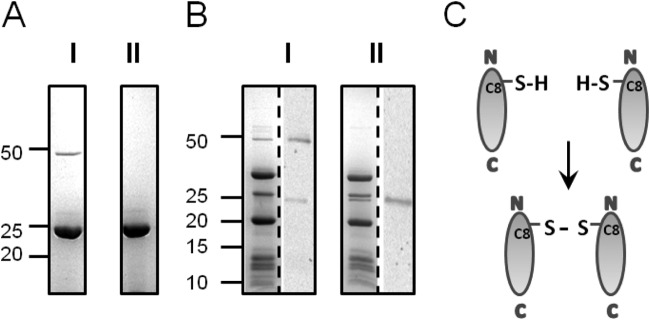
FIG 2.

VP11 dimer formation. (A) Coomassie blue-stained SDS-15% polyacrylamide gel of purified recombinant VP11 boiled in SDS sample buffer with 1% β-mercaptoethanol (I) or 5% β-mercaptoethanol (II). (B) P23-77 virus particles prepared with 1% β-mercaptoethanol (I) and 5% β-mercaptoethanol (II) as for panel A. The protein pattern of a Coomassie-stained gel is shown on the left side of each gel, and Western blots incubated with VP11 polyclonal antibody are shown on the right side. (A and B) Protein sizes are indicated in kilodaltons according to the All Blue Precision Plus protein standard (Bio-Rad). (C) Schematic presentation of intermolecular disulfide bond formation between cysteine residues at the N terminus of VP11.
