
FIG 7.
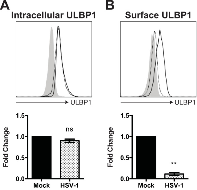
HSV-1 downregulates ULBP1 on the cell surface but not intracellularly. 293T cells were infected with HSV-1 (MOI of 3) or mock infected. Twenty-four hours postinfection, cells were harvested and stained with anti-ULBP1 antibody for analysis by flow cytometry. Levels of intracellular (A) and surface (B) ULBP1 expression were compared between mock (continuous black line) and HSV-1 (dotted black line) infections. Isotype control antibody staining is indicated by the gray-filled histogram. One representative histogram from at least two independent experiments is shown. The relative MFI fold change over mock infection in ULBP1 expression (less respective isotypes) is presented below each correlating histogram as the mean ± SEM of data from biological replicates (A, n = 2; B, n = 3). Statistical significance was established by two-tailed paired Student's t test. ns, not significant; **, P < 0.01.