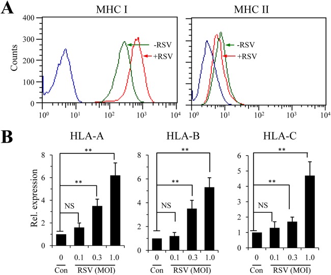
FIG 1.
Determination of MHC-I and MHC-II expression in RSV-infected A549 airway epithelial cells. (A) Surface expression of MHC-I and MHC-II by flow cytometry study. The A549 cells of a human alveolar basal epithelial cell line were infected with RSV at an MOI of 1.0 for 36 h. Surface expression of MHC-I or MHC-II was stained for with commercial antibody kits and determined by FACS. Values on the x axes are fluorescence intensities of APC-MHC I and FITC-MHC II, respectively. Blue lines indicate results for the idiotype controls. The experiment was performed independently 3 times. (B) Determination of MHC-I molecules by quantitative PCR. A549 cells were left uninfected (Con) or infected with various amounts of RSV for 24 h. Expression of the HLA-A, HLA-B, and HLA-C genes was determined by qPCR. The results are representative of three independent experiments with duplicate samples. The data are presented as means and standard deviations (SD) of duplicate samples. An unpaired t test was performed for statistical analysis. NS, no significance; **, P ≤ 0.01.

