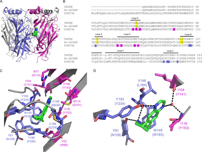
Figure 1.
Varenicline bound to 5-HTBP. (A) Location of varenicline (green) at the interface between two subunits in the orthosteric 5-HTBP binding site. (B) Alignment of 5-HTBP, AChBP, and the extracellular domain of the 5-HT3A receptor subunit showing the approximate location of the A–E binding loops. The residues mutated in this study are highlighted in purple, and the residues that differ between 5-HTBP and AChBP in yellow. (C) The 5-HTBP binding pocket showing the orientation of varenicline (green) and nearby residues on the principal (blue) and complementary (magenta) faces. The corresponding 5-HT3 receptor residues are in parentheses. (D) Hydrogen bonds are present between varenicline and residues Y91, Y193, and W145 on the principal face and residues I104 and I116 on the complementary face via a water molecule.

