
Fig. 2.
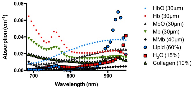
Chromophores used in the fitting routine to approximate near-infrared absorption spectra in cardiac tissues. All spectra were obtained from previously published literature. Water (H2O)a; oxyhemoglobin (HbO2)a; deoxyhemoglobin (Hb)a; lipidb; reduced oxymyoglobin (Mb)b; met-myoglobin (met-Mb)b; oxymyoglobin (MbO2)b; and collagenc; a-Obtained from Jacques et al.; b-Obtained from Bowen et. al.; c-Obtained from Taroni et. al.