
Figure 2.
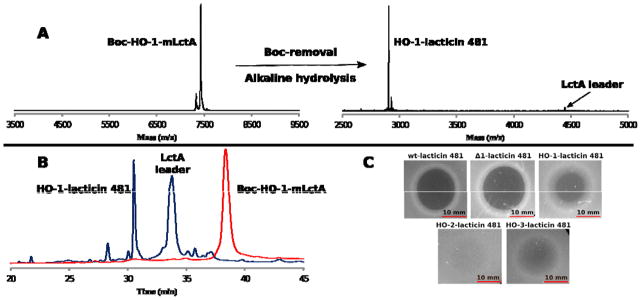
A) MALDI-TOF MS spectra depicting fully modified LctA containing Boc-HO-1 at position 1 (Boc-HO-1-mLctA), and the Boc-removal from and alkaline hydrolysis of Boc-HO-1-mLctA yielding HO-1-lacticin 481 and LctA leader peptide. The leader peptide ionizes poorly in the mass spectrum, but is seen clearly by HPLC. Boc-HO-1-mLctA ([M+H]+; calc’d = 7432 Da; observed = 7428 Da). HO-1-lacticin 481 ([M+H]+; calc’d = 2901.3 Da; observed = 2901.3 Da; monoisotopic mass spectrum). B) Analytical RP-HPLC trace of HO-1-mLctA before (red) and after (blue) Boc removal and alkaline hydrolysis. C) Antimicrobial assays of lacticin 481 and analogues against L. lactis HP. A total of 10 μL of 12.5 μM compound solution was added to each spot. Plates were incubated at 30 °C for 18 h.