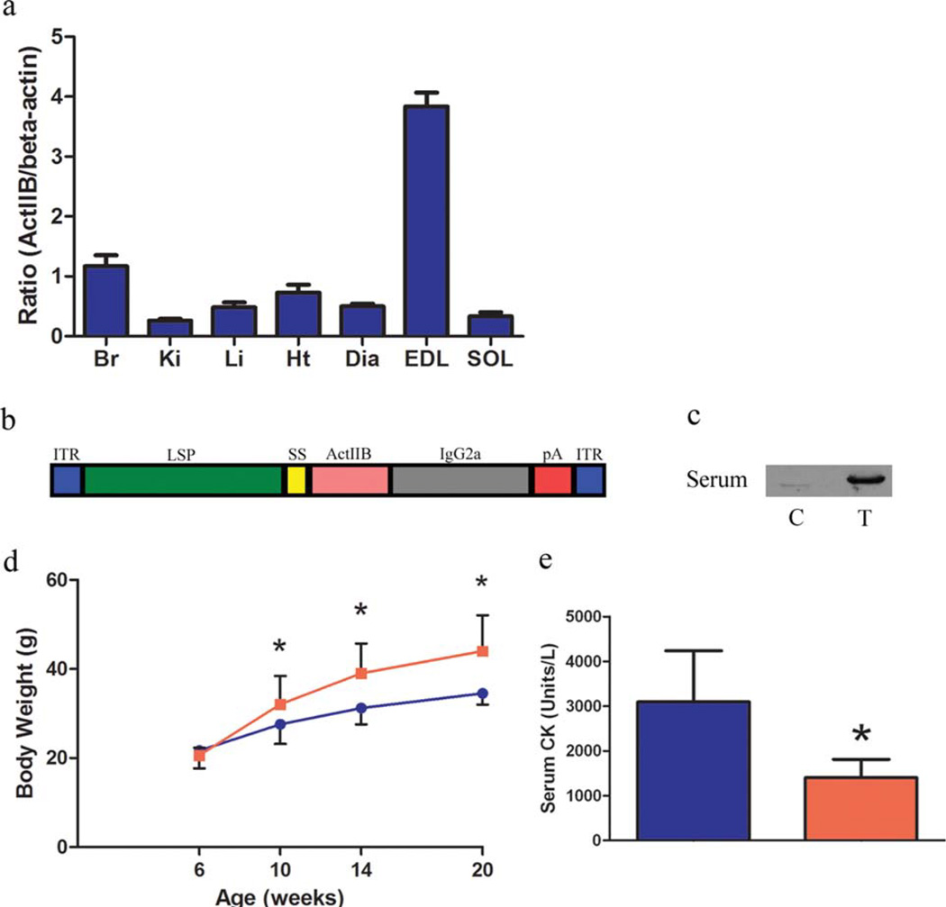
FIGURE 1.
Expression of soluble activin IIB receptor in mdx mice. (a) Tissue distribution of the activin IIB receptor assessed by immunoblotting. (b) A schematic of the AAV construct used in this study. (c) Six-week-old mdx mice (n = 5 control, n = 5 treated) were injected intraperitoneally with 1E12 genome copies of AAV 2/8 LSP.sActIIBr or saline. Immunoblotting of control (C) and treated (T) serum samples 1 month after injection demonstrates the presence of circulating sActIIBr. (d) Significantly increased body weight was noted in the mdx/sActIIBr group (shown in red) in comparison to control mdx mice (shown in blue) from 1 month post-injection until the conclusion of the study. (e) Activin receptor blockade significantly reduced serum CK from 3095 ± 1139 U/L in controls (blue bar) to 1404 ± 409 U/L in treated mice (red bar). Br, brain; Ki, kidney; Li, liver; Ht, heart; Dia, diaphragm; EDL, extensor digitorum longus; SOL, soleus. *P < 0.05. [Color figure can be viewed in the online issue, which is available at wileyonlinelibrary.com.]

