
FIG. 1.
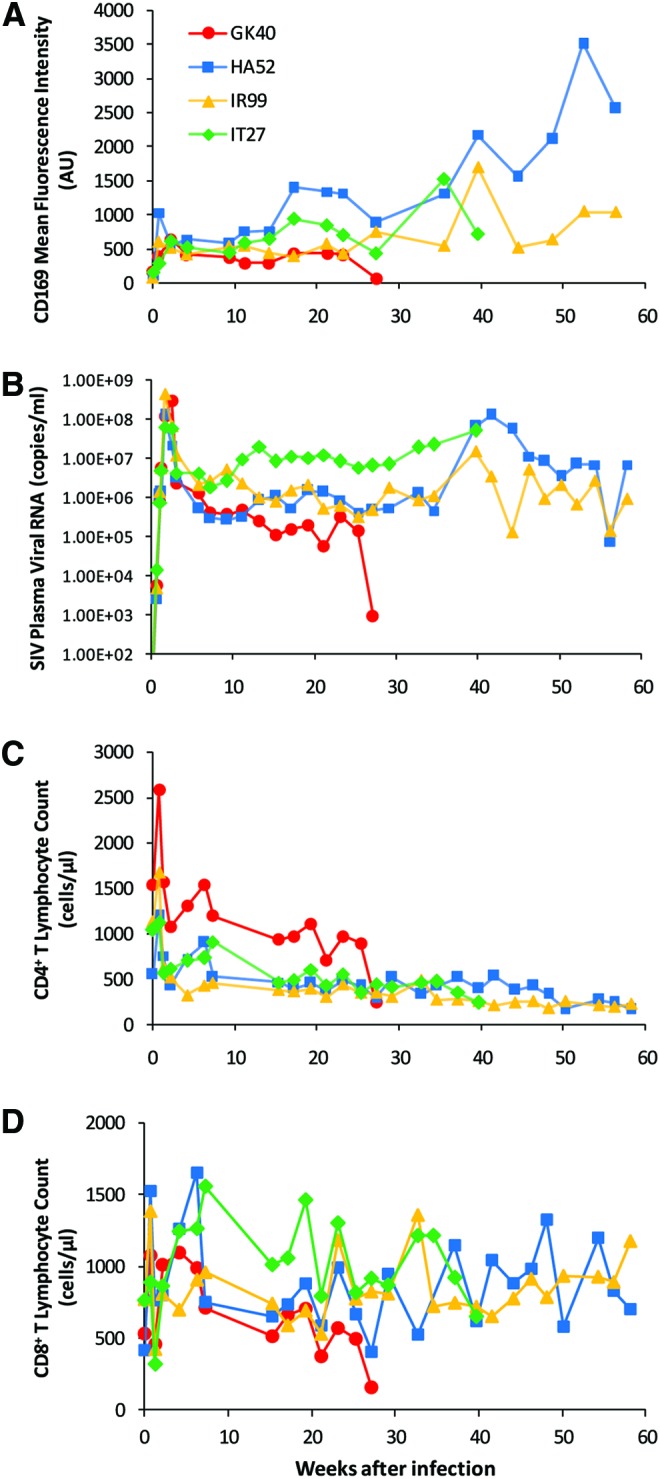
Increased expression of CD169 on monocytes during the course of SIV infection. Four adult rhesus macaques were infected with SIVmac251. (A) Monocyte CD169 mean fluorescence intensities (arbitrary units, AU) measured by flow cytometry in individual macaques after infection are plotted over time. After being gated by their forward and side scatter properties, monocytes were further defined as CD14+HLA-DR+ cells that are negative for CD3, CD8, and CD20. The mean fluorescence intensity of CD169 on total monocytes was normalized to the corresponding isotype control. (B) Plasma SIV viral loads in individual macaques after infection are shown. (C) Absolute counts of CD4+ T lymphocytes and (D) absolute counts of CD8+ T lymphocytes in individual macaques after infection are shown. Note: one animal (GK40) died early at around week 27 pi. Color images available online at www.liebertpub.com/aid