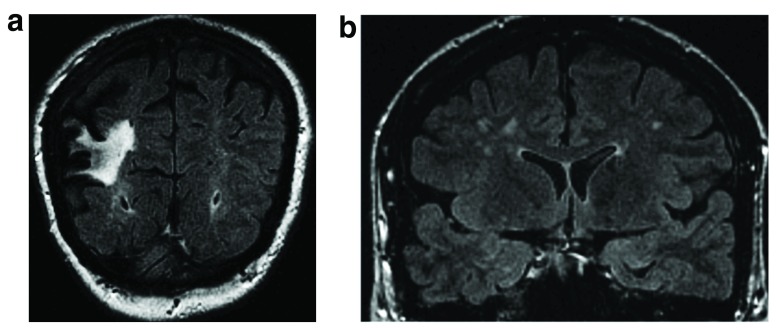
Figure 1a–b. Coronal FLAIR images demonstrate both large and small vessel disease.
1a. Large hyperintense cortico-subcortical area consistent with a chronic stroke involving the right middle cerebral artery territory. 1b. Focal bilateral white matter hyperintensities reflecting small vessel disease. Figure origin: Department of Radiology, Hospital Clinic Barcelona.

