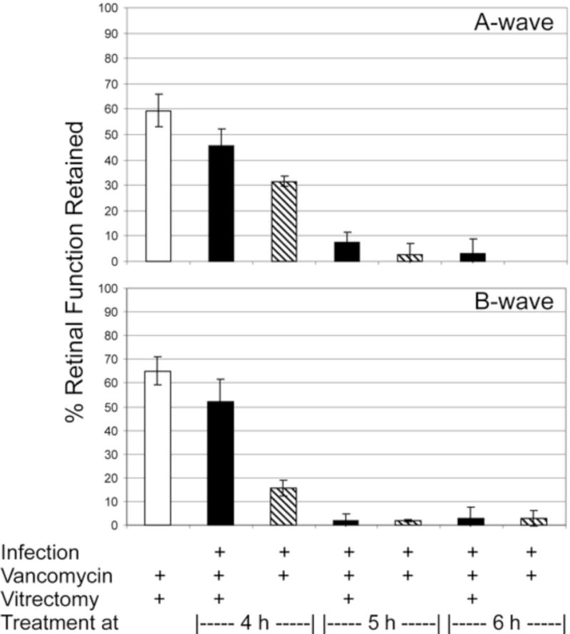
Figure 1.

Retinal function analysis following treatment of experimental B. cereus endophthalmitis with vitrectomy and vancomycin. Eyes were infected with B. cereus and treated with vancomycin (1%) and vitrectomy or vancomycin alone at various times postinfection. The control group included uninfected eyes treated with vitrectomy and vancomycin. Eyes were analyzed by electroretinography at 36 h or 48 h postinfection postinfection. Values represent the mean ± SEM of N≥5 eyes per group.
