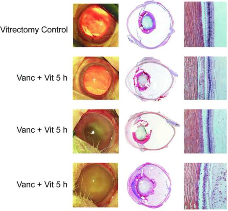
Figure 2.

Photography and histology of eyes following treatment of experimental B. cereus endophthalmitis with vitrectomy and vancomycin (1%) at various times postinfection. The control group included uninfected eyes treated with vitrectomy + vancomycin. Eyes were photographed, then harvested for histology and hematoxylin and eosin staining. Figures are representative of N=3 eyes per group. Vanc = vancomycin, Vit = vitrectomy. Magnification of retina sections, 100×.
