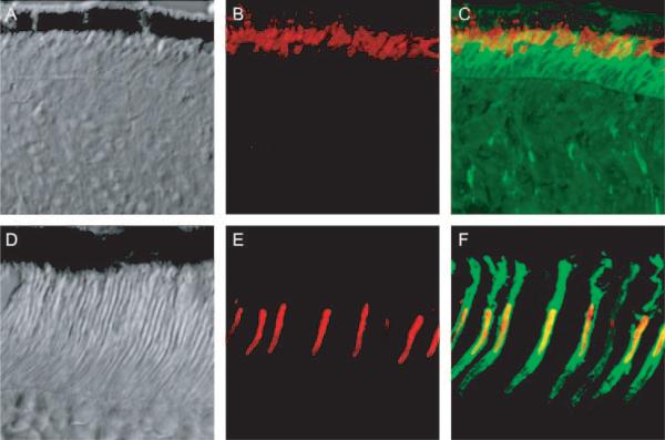
Figure 3.
(A–C) Images of a frozen section of Nrl−/− retina taken with differential interference contrast optics (A), with fluorescent immuno-staining of MUV, the mouse ultraviolet pigment (B, red), and with overlaid fluorescent immunostaining of MUV and PNA binding (green) (C). (D–F) Images of a frozen section of a WT retina stained and presented in the same format as in (A–C). Images were made with a confocal microscope and represent 50 × 50-μm regions of the two retinas.

