
Figure 4.
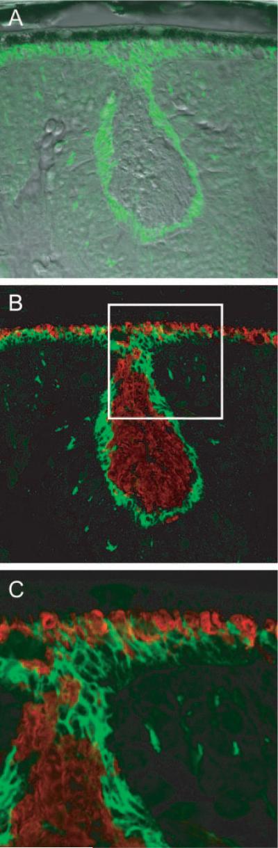
Confocal images of a rosette in the ONL of the Nrl−/− retina. (A) Differential interference contrast image with overlaid image taken of PNA labeling (green). (B) Same section as in (A), but merging immunostaining of MUV (red) and PNA labeling. MUV is seen to fill the center portion of the rosette; the white square is 50 × 50 μm. (C) Magnified image of the portion of (B) highlighted by the white square, showing details of a portion of the rosette. Note how the PNA labeling of the border of the rosette merges with the PNA-stained inner segment layer of the cones that are in contact with the RPE.