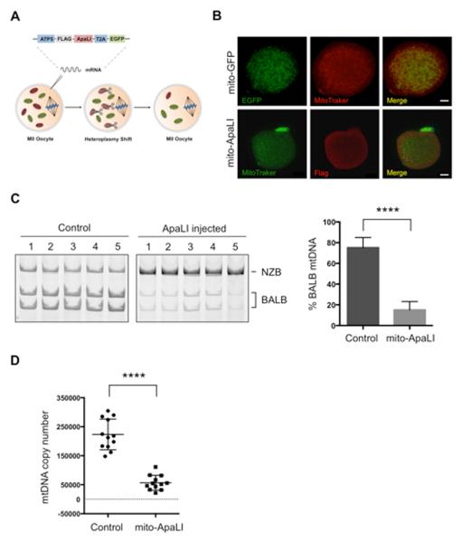
Figure 1. Heteroplasmy shift in NZB/BALB MII oocytes using mito-ApaLI.
(A) Injection of mito-ApaLI mRNA in oocytes for induction of heteroplasmy shift.
(B) Mitochondrial co-localization of mito-GFP and mito-ApaLI with Mitotracker in injected oocytes by immunofluorescence. Scale bars, 10μm.
(C) RFLP analysis and quantification of mtDNA heteroplasmy in control and mito-ApaLI injected MII oocytes after 48 h (Control n=16; mito-ApaLI n=12). Representative gel.
(D) Quantification of mtDNA copy number by qPCR in control and mito-ApaLI injected oocytes MII after 48 h (Control n=12; mito-ApaLI n=12).
Error bars represent ± SEM. ****p<0.0001. See also Figure S1.

