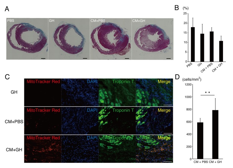
Fig 3. GH enhanced engraftment of CM.
(A-B) Cardiac sections were stained with azan to evaluate the infarcted area. The CM+GH group tended to have a smaller infarcted area. Bars are 1 mm. (C) CM were prestained with MitoTracker-Red and sections were co-stained with cardiac troponin T and DAPI. In the GH group, there were no red signals in infarcted hearts. In the CM+PBS group, few CM were engrafted in the infarcted area. In the CM+GH group, more CM remained in the infarcted area. Bars are 100 μm. (D) The number of engrafted CM was increased significantly when transplanted with GH compared with CM transplanted with PBS. (** P<0.01)

