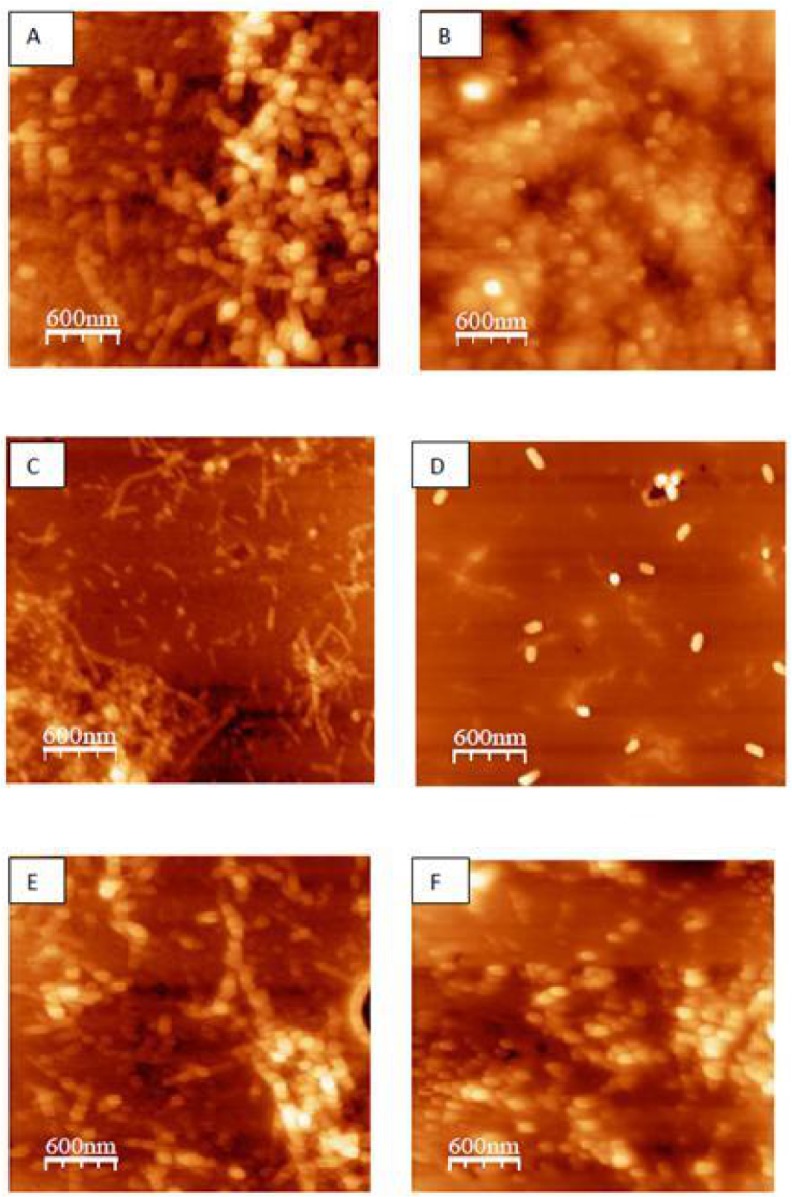
Fig 3. AFM images of different β-Lg samples incubated at 80°C and pH 2 for 24 h.

(A) β-Lg alone, (B) β-Lg in the presence of curcumin, (C) β-Lg in the presence of AFM1 (1 ppb), (D) β-Lg in the presence of AFM1 (1 ppb) and curcumin, (E) β-Lg in the presence of Pb2+ (0.2 ppm), and (F) β-Lg in the presence of Pb2+ (0.2 ppm) and curcumin.
