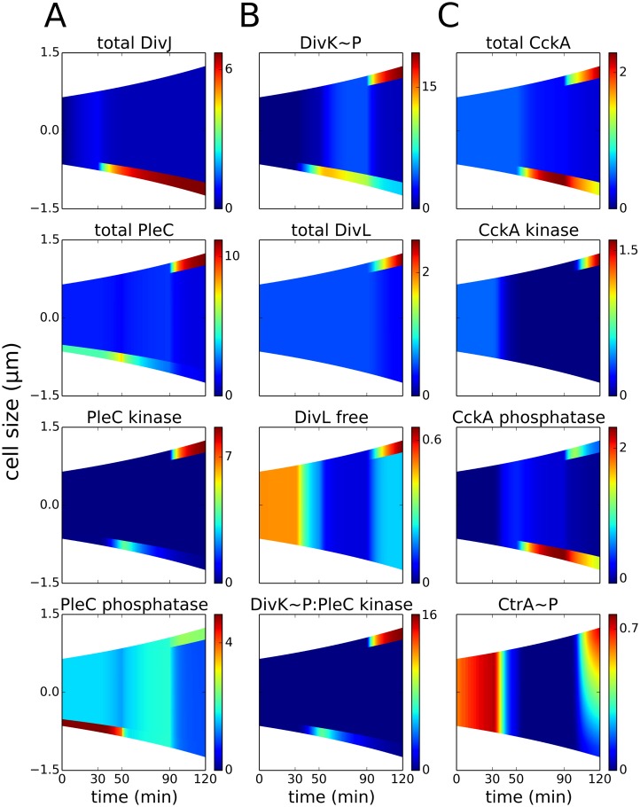
Fig 3. Co-localization of PleC kinase and DivL in the early predivisional cell is required for DivL reactivation.
Spatiotemporal distributions of proteins during the cell cycle (prior to cytokinesis at t = 120 min). Color indicates concentration gradients from minimum (blue) to maximum (red). (A) DivJ is localized at the old pole (t = 30–120 min). The location of PleC is shifted from the old pole (t = 0–50 min) to the new pole of the predivisional cell (t = 90–150 min). Following DivJ localization, the function of PleC changes from a phosphatase to a kinase. (B) Upon phosphorylation, DivK localizes to the poles of the cell. Despite the presence of DivK~P at the new pole of the predivisional cell, DivL is present in the free form (unbound to DivK~P) because DivL co-localizes with PleC kinase and PleC kinase sequesters DivK~P, preventing it from binding to DivL. (C) CckA is uniformly distributed in the swarmer stage and localized at both poles in the predivisional stage. Reactivation of DivL at the new pole results in new-pole CckA becoming a kinase, while old-pole CckA remains a phosphatase. Consequently, the late predivisional cell establishes a gradient of CtrA~P along its length from high at the new pole to low at the old pole.

