
Figure 4. New Hepatocytes Are Not Labeled on AAV Serum Transfusion.
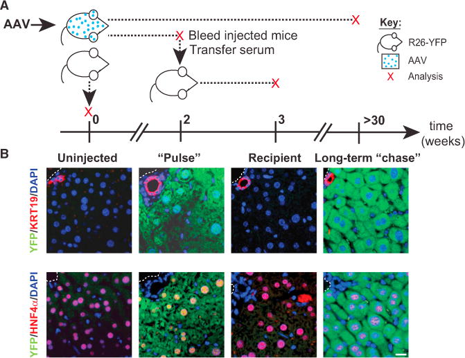
(A) Schematic showing the timing of infection, serum transfer, and analysis of injected R26YFP mice; each red “x” indicates a sampling point. AAV8-TBG-Cre-injected mice were analyzed either 2 weeks (“Pulse”) or >30 weeks after injection (Long-term “chase”). Prior to analysis at 2 weeks, mice were bled, and serum was injected into naive, uninfected R26YFP mice whose livers were assessed after 1 week (Recipient). Uninfected mice served as a control (Uninjected).
(B) Images showing immunoflurescence staining for YFP (green) and Krt19 or HNF4α (red) in livers from each of the four groups analyzed.