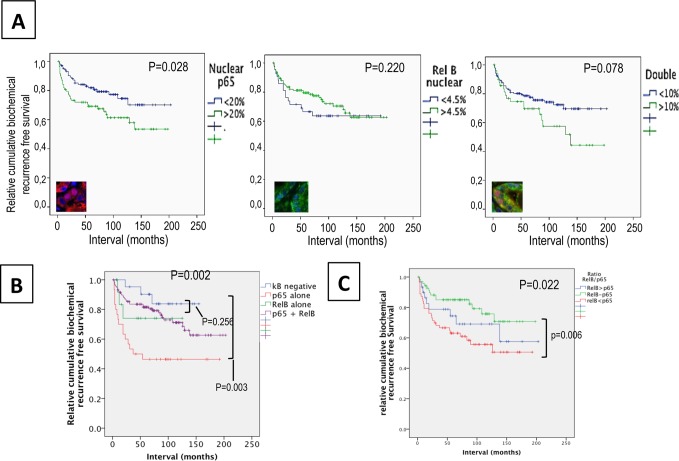
Fig 3. Association between nuclear p65 and/or RelB and biochemical recurrence in prostate cancerpatients.
Kaplan–Meier biochemical recurrence-free survival curves A. High (>20%) and low (<20%) frequency of nuclear p65 in the epithelia of cancer tissues. B. High (>5%) and low (<5%) frequency of nuclear RelB in the epithelia of cancer tissues. Significance (p) is indicated by log rank. C. Double nuclear epithelial staining of p65 and RelB. D. Epithelial and nuclear staining of p65 and RelB. The κB negative variable represents patients without positive tissue cores for p65 and RelB. The variable p65 alone or RelB alone represents patients positive for only nuclear p65 or nuclear RelB. The variable p65 + RelB are patients with presence of both nuclear subunits in the same core. E. Analysis of the epithelial proportion of nuclear p65 to RelB in tissue cores within both p65 and RelB staining. The ratio p65/RelB represents the frequency of p65 to the frequency of RelB. The variable p65 > RelB represents a ratio of at least 2 fold more p65 than RelB; p65-RelB represents a ratio between 0.50 and 2 fold, and RelB>p65 represents a ratio of at least 2 fold more RelB than p65.

