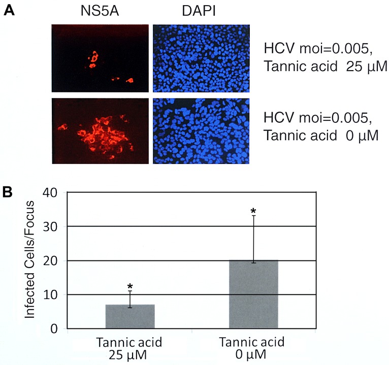
Fig 6. Effect of tannic acid on cell-to-cell spread of HCV.
(A) The effect of tannic acid on cell-to-cell spread of HCV as measured by viral protein staining. Huh7.5 cells were infected with HCV using an MOI of 0.005 at 37°C. The inoculum was removed two hours post infection and replaced with a 1% agarose medium overlay containing 25 μM tannic acid. Controls had no tannic acid. Cells were incubated at 37°C for 72 hours as described in Materials and Methods. Infected cells were labeled by indirect immuno-fluorescence using an anti-HCV NS5A monoclonal antibody (red), nuclei were stained with DAPI (blue) and photographs were taken with a fluorescence microscope as described in Materials and Methods. (B) The mean and standard deviation of infected cells/focus was determined by visual counting of 30 foci present on randomly selected fields of cover slips for each condition. P-values were calculated using the Student’s ttest (asterisk indicates P = 0.000019). Experiments were performed three times and representative examples are shown in A and B.

