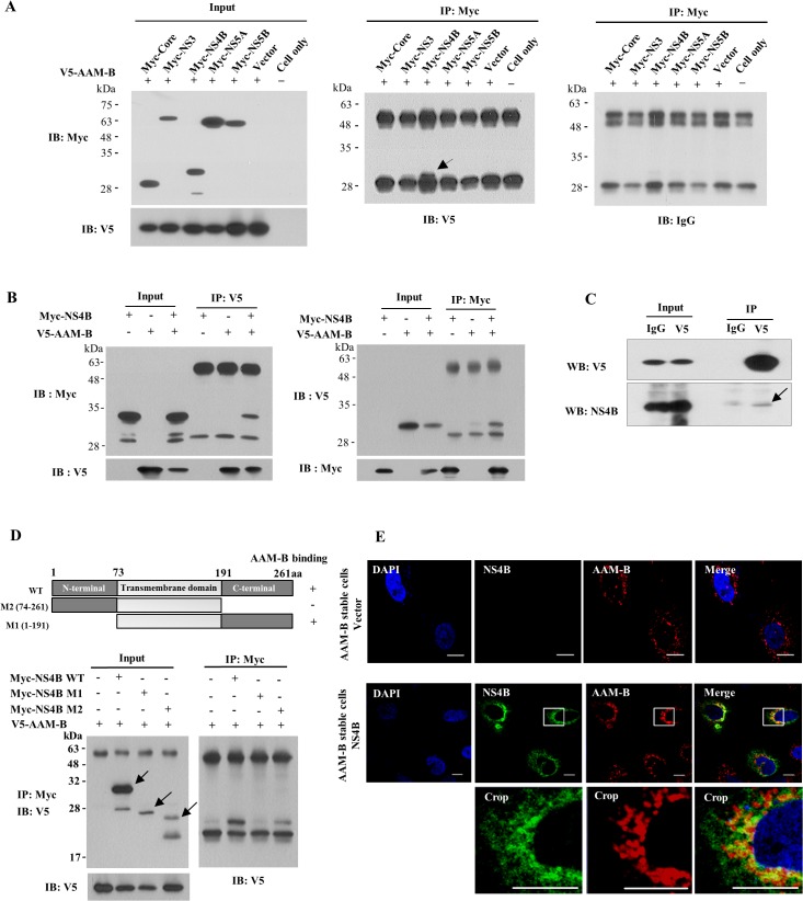
Fig 5. AAM-B interacts with HCV NS4B protein.
(A) HEK293T cells were cotransfected with V5-tagged AAM-B plasmid and Myc-tagged HCV protein as indicated. Protein expressions of input plasmids were confirmed by immunoblot analysis using either anti-Myc antibody (Left, upper) or anti-V5 antibody (Left, lower). (Right) Total cell lysates were immunoprecipitated with anti-Myc antibody and then bound proteins were immunoblotted with anti-V5 antibody. The arrow indicates the interacting band. (B) (Left) HEK293T cells were cotransfected with V5-tagged AAM-B plasmid and Myc-tagged NS4B (genotype 1b) expressing plasmid. At 48 h after transfection, total cell lysates were immunoprecipitated with anti-V5 antibody and then bound proteins were immunoblotted with anti-Myc antibody. (Right) The same cell lysates were immunoprecipitated with anti-Myc antibody and bound proteins were immunoblotted with anti-V5 antibody. (C) Huh7 cells harboring subgenomic replicon derived from genotype 1b were transfected with V5-tagged AAM-B expression plasmid. Total cell lysates were immunoprecipitated with rabbit anti-V5 antibody and then bound proteins were immunoblotted with either mouse anti-V5 antibody (left) or mouse anti-NS4B (right). The arrow indicates the interacting band. (D) (Upper) Schematic representation of both wild-type and mutant constructs of NS4B protein. (Lower) HEK293T cells were cotransfected with V5-tagged AAM-B and Myc-tagged mutant constructs of NS4B. Total cell lysates harvested at 48 h after transfection were immunoprecipitated with anti-Myc antibody and bound proteins were immunoblotted with anti-V5 antibody. Both wild-type and mutant protein expressions of NS4B were verified by immunoprecipitation and immunoblot analysis using anti-Myc antibody (arrows). (E) AAM-B stable cells were transiently transfected with either vector or NS4B expression plasmid. At 48 h after transfection, cells were fixed in 4% paraformaldehyde. Cells were washed twice with PBS and then incubated with either anti-NS4B or anti-AAM-B antibodies for 1 h in room temperature. Immunofluorescence staining was performed by using either FITC-conjugated or TRICT-conjugated secondary antibodies. The inset in each panel shows enlarged image of the area marked in a white square. Bars, 10 μm.

