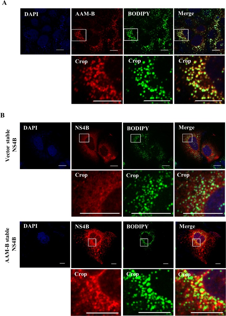
Fig 6. AAM-B colocalizes with NS4B in the proximity to the lipid droplets.
(A) AAM-B stable cells fixed with paraformaldehyde were incubated with anti-V5 antibody to detect AAM-B and BODIPY. AAM-B colocalized with LD in the cytoplasm. Bars, 10 μm. (B) Either vector stable or AAM-B stable cells were transiently transfected with NS4B plasmid. At 48 h after transfection, cells were fixed in 4% paraformaldehyde and incubated with anti-NS4B antibody and BODIPY for 1 h at 37°C. Samples were analyzed for immunofluorescence staining using the LSM 700 laser confocal microscopy system. Cells were counterstained with DAPI to label nuclei. The insets in the panels show enlarged views of the areas marked in white squares. Bars, 10 μm.

