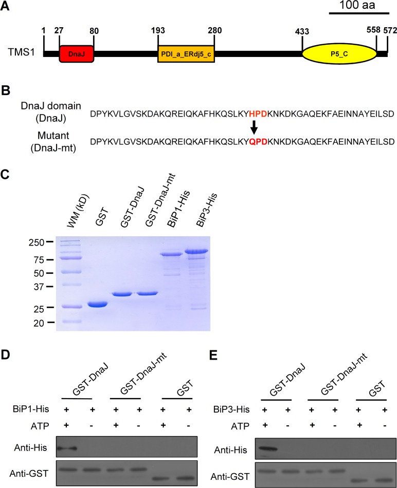
Fig 3. The DnaJ domain of TMS1 could bind to BiP1 and BiP3 in vitro.
(A) A schematic structure of the TMS1 protein, indicating the position of the DnaJ domain in the TMS1. (B) The amino acid sequence of the TMS1 DnaJ Domain and the loss-of-function mutant DnaJ Domain. The red characters indicate the position of the HPD motif and the mutation site in the mutant protein. (C) The quality assay for the purified proteins. (D) The in vitro GST pull-down demonstrated that DnaJ domain of TMS1 could bind to BiP1 when supplied with ATPs. (E) The in vitro GST pull-down demonstrated that DnaJ domain of TMS1 could bind to BiP3 when supplied with ATPs. 20-fold dilutions of the protein samples were used for the anti-GST assays in (D) and (E). WM, molecular weight marker; kD, kilodaltons.

