FIGURE 3.
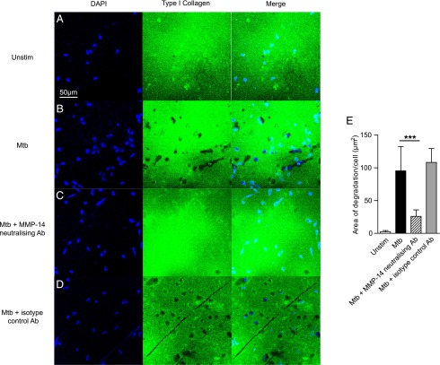
M. tuberculosis–driven collagen degradation is MT1-MMP–dependent. (A and B) Human monocytes were seeded on FITC-conjugated type I collagen and infected with M. tuberculosis. Collagen degradation was analyzed by immunofluorescent microscopy. Blue is DAPI nuclear stain; green is type I collagen. M. tuberculosis infection causes greater collagen degradation. (C) MT1-MMP activity was neutralized with an inhibitory anti-MT1-MMP Ab (MAB3328, Millipore) at 10 μg/ml. MT1-MMP inhibition inhibited areas of collagen breakdown. (D) An isotype control Ab did not inhibit collagen degradation. (E) Fluorescence quantitation confirms collagen degradation is MT1-MMP–dependent. Data are from a single experiment and are representative of three independent experiments. Mean values ± SD are shown. ***p < 0.001 by one-way ANOVA with a Tukey post hoc test.

