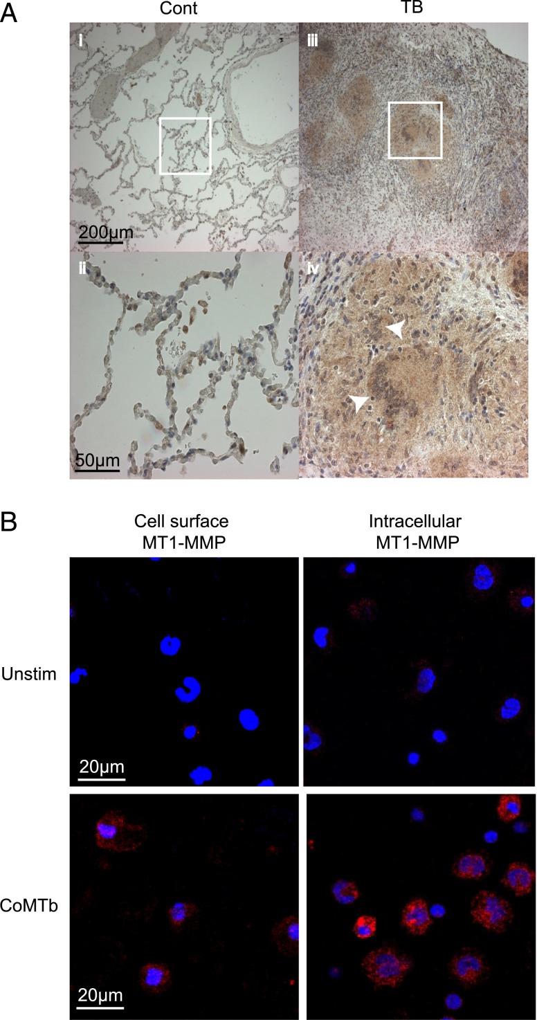
FIGURE 4.
MT1-MMP is expressed in TB granulomas from patients and is driven by monocyte–monocyte networks. (A) Lung biopsies from five patients with culture-proven M. tuberculosis infection and control tissue from uninvolved lung parenchyma of patients with lung cancer were immunostained for MT1-MMP. In control biopsies, MT1-MMP immunostaining is present only in alveolar macrophages (i and ii). In TB, MT1-MMP immunoreactivity is present throughout the granuloma, expressed by both giant cells and epithelioid macrophages (iii and iv). White arrows indicate Langerhans multinucleate giant cells, surrounded by epithelioid macrophages. (B) Monocytes were stimulated with CoMTb to model intercellular networks. MT1-MMP expression was measured in unpermeabalized and permeabalized monocytes by immunofluorescent staining and microscopy. Blue is DAPI nuclear stain, magenta is MT1-MMP. CoMTb stimulation increased both surface and intracellular MT1-MMP expression at 24 h. Images are representative of three independent experiments.

