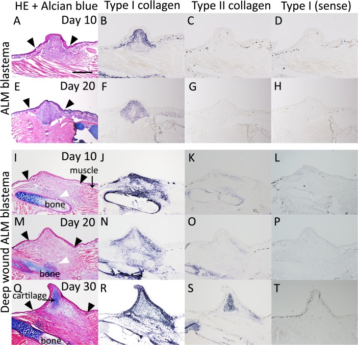
Fig 8. Type I and type II collagen expression patterns in Xenopus ALM blastemas.
(A-H) The Xenopus ALM blastema. (A-D) The Xenopus ALM blastema at 10 days postoperation. (A) HE and Alcian blue staining. (B) Type I collagen expression. (C) Type II collagen expression. (E-H) The Xenopus ALM with deep wound blastema at 20 days postoperation. (E) HE and Alcian blue staining. (F) Type I collagen expression. (G) Type II collagen expression. (I-T) The Xenopus ALM with deep wound blastema. (I-L) The Xenopus ALM with deep wound blastema at 10 days postoperation. (I) HE and Alcian blue staining. (J) Type I collagen expression. (K) Type II collagen expression. (M-P) The Xenopus ALM with deep wound blastema at 20 days postoperation. (M) HE and Alcian blue staining. (N) Type I collagen expression. (O) Type II collagen expression. (Q-T) The Xenopus ALM with deep wound blastema at 30 days postoperation. (Q) HE and Alcian blue staining. (R) Type I collagen expression. (S) Type II collagen expression. (D, H, L, P, T) Control of in situ hybridization experiments. Sense probe of type I collagen. All are shown at the same magnification. Scale bar is 500 μm. Black arrowheads indicate wound line. White arrowheads indicate bone cracked region.

