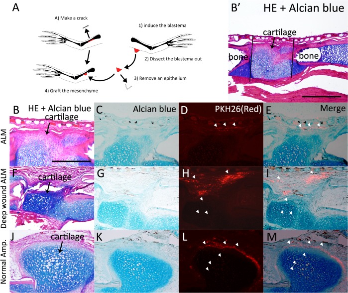
Fig 9. Xenopus ALM blastema cells do not have cartilaginous differentiation capacity.
(A) The scheme of the experiment. (B-E) Xenopus ALM blastema cells were grafted to the bone wound site. (B) HE and Alcian blue staining. B’ is a lower magnification image of B. Black lines indicate bone crack area. (C) Alcian blue staining. The cartilaginous callus was visualized by Alcian blue stain. (D, E) Grafted cells were PKH26-positive (red). PKH26-positive cells were not observed in cartilaginous callus. White arrow heads indicate PKH26-positive cells. (F-I) Deep wound ALM blastema cells were grafted to the bone wound site. (F) HE and Alcian blue staining. (G) Alcian blue staining. (H, I) Grafted cells were observed in the cartilaginous callus. (J-M) Control experiment. Normal blastema cells were grafted to the bone wound site. (J) HE and Alcian blue staining. (K) Alcian blue staining. (L, M) Grafted cells were observed in the cartilaginous callus. B-M are shown at the same magnification. Scale bar in B is 200 μm. Scale bar in B’ is 500 μm.

