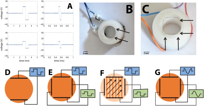
Fig 1. Apparatus used to stimulate neuronal cultures with external electric fields.
(A) Waveforms of the measured applied voltage between the electrodes (upper row) and within the fluid (lower row), approximately at the center of the experimental cell where the culture is located. Left column is for pulse durations of 1ms and the right is for durations of 100 μs. (B) The plastic insert with one pair of electrodes used to stimulate the neuronal culture. The electrode wires are indicated in the picture by arrows. A plastic rim extending into the medium locates the two parallel platinum wire electrodes about 1 mm above the culture. (C) The device used to stimulate the neuronal culture with two orthogonal pairs of electrodes. A plastic rim holds two pairs of platinum electrodes about 1mm above the culture, which are denoted in the picture by arrows. (D) Sketch of the electrical circuit for a pair of electrodes that is driven by a single square pulse with varying durations. The electric field created between these electrodes is used to stimulate a neuronal network cultured on a glass coverslip. (E, F) Sketch of the electrical circuit for two pairs of electrodes that are fed with separate single square pulses with varying durations (synchronized but with no common ground). Changing the relative amplitudes changes the orientation of the electrical field, which is used to directionally stimulate a neuronal network cultured on a cover slip. (G) Sketch of the electrical circuit for obtaining a rotating electric field. Two pairs of electrodes are each fed with one cycle of sine or of cosine voltage pulses (i.e. two signals with the same amplitude but phase shifted by π/2).

