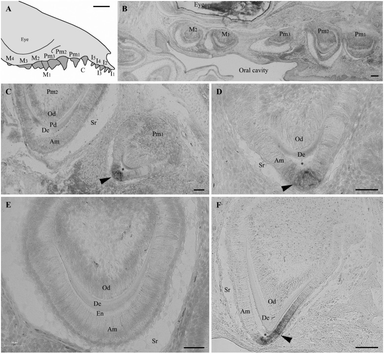
Fig 2. Amelotin gene expression during amelogenesis in Monodelphis domestica teeth.
(A) Schematical drawing of the upper jaw of a Monodelphis domestica neonate (lateral view). (B-F) In situ hybridization of sagittal tooth sections in the upper jaw of a 16 days neonate using AMTN probe. (B) Low magnification of a section showing five developing teeth. No labelling of AMTN transcripts are observed although the teeth are already formed and show well-differentiated ameloblasts. Enamel matrix is present but not yet undergoing maturation process, at least in this section level. (C) Section of premolars 2 (left) and 1 (right). In the less developed premolar 2 (secretory stage), the ameloblasts, which are well-differentiated, display Tomes' processes and are facing recently formed enamel matrix, are not labelled. In contrast, in premolar 1, labelled AMTN transcripts (arrowhead) are observed in a few ameloblasts located at the tooth tip and facing the mineralized enamel (asterisk). (D) Higher magnification of Pm1 in (C). Mature enamel (asterisk) facing labelled ameloblasts (arrowhead) was removed during the demineralization process. (E) Similar stage as Pm2 in (C). The secretory-stage ameloblasts display Tomes' processes but do not express AMTN. Immature enamel matrix is present between the ameloblasts and the dentin layer. (F) In the upper region of premolar 1 AMTN expression is only located in the short, maturation-stage amelobasts facing the well-mineralized enamel (asterisk). The ameloblasts facing the immature enamel matrix (on the left) are not labelled. Am: ameloblasts; De: dentin; En: enamel matrix; Od: odontoblasts; Pd: predentin; Sr: stellate reticulum; *: enamel space. C: canine; I: incisor; M: molar; Pm: premolar. Scale bars: A = 2 mm; B, F = 100 μm; C, D, E = 50 μm.

