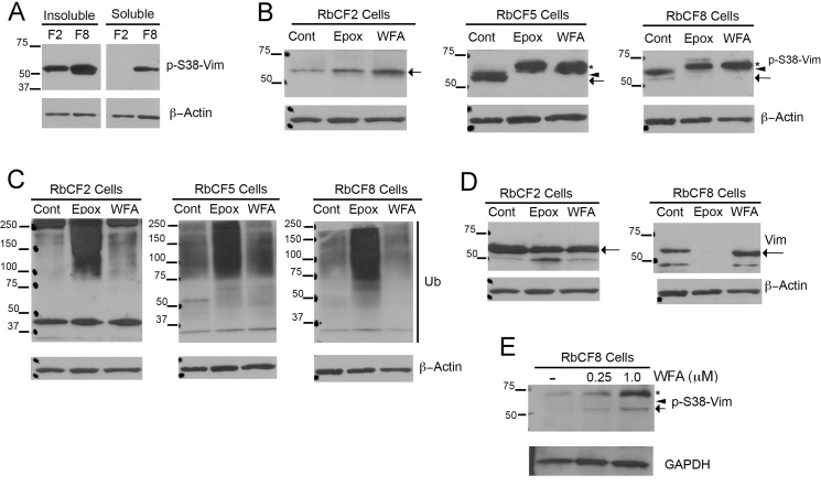
Fig 5. Hyperphosphorylation of soluble pSer38vim by WFA is cell passage dependent.
(A) RbCF2 and RbCF8 cells were cultured as in Fig 1 and plated for 1 h. Equal amounts of soluble and insoluble proteins were fractionated by SDS-PAGE and blots were probed with anti-pSer38vim and β-actin antibodies. (B) RbCF2, RbCF5 and RbCF8 cells were cultured as in Fig 1, plated for 30 min and then treated in presence or absence of WFA or Epox for 30 min. Equal amount of soluble proteins were fractionated by SDS-PAGE and blots were probed with anti-pSer38vim and β-actin antibodies. Asterisk marks the 67-kDa high molecular weight pSer38vim hyperphosphorylated species, and the arrow and arrowheads represent the 57 kDa and 61 kDa pSer38vim bands, respectively. (C) Western blots of RbCF2, RbCF5 and RbCF8 cells re-probed with anti-ubiquitin antibody. (D) Western blots comparing sVim expression in RbCF2 and RbCF8 cells cultured as in Fig 1 and treated with Epox or WFA as above. The arrow indicates the major vimentin 57 kDa species. (E) Western blots of RbCF8 cells treated with WFA in suspension culture. Serum-starved cells were trypsinized and transferred into non-adhesive petri dishes in 10% serum-containing medium in presence or absence of WFA for 1 h. Equal amount of soluble proteins were fractionated by SDS-PAGE and blots were probed with anti-pSer38vim and β-actin antibodies. The arrow points to the 57 kDa species, arrowhead to 61 kDa species and asterisk to the 67 kDa species.

