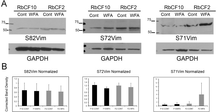
Fig 6. Vimentin phosphorylation at other serine residues.
RbCF2 and RbCF8 cells were cultured as in Fig 1, plated for 30 min followed by treatment in presence or absence of WFA (1 μM) for 1 h. (A) Equal amount of soluble proteins were fractionated by SDS-PAGE and blots were probed with anti-S82vimentin (clone MO82), anti-S71vimentin (clone TM71) and anti-S72vimentin (clone TM72), respectively. Blots were then stripped and re-probed for loading controls. (B) Densitometric quantification of repeat experiments (n = 3; see also S1 and S2 Figs) of S82Vim, S72Vim and S71Vim normalized to loading controls, using NIH ImageJ software.

