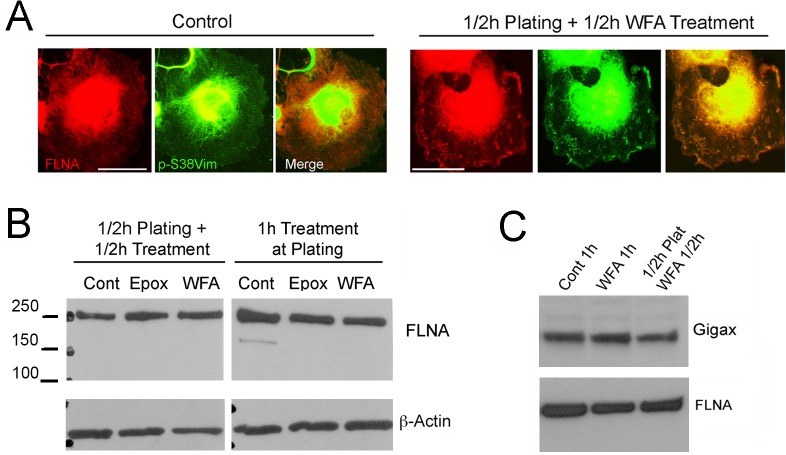
Fig 7. WFA alters Filamin A organization but not its expression.
(A) RbCF8 cells were cultured as in Fig 1 in presence or absence of WFA or Epox for 30 min. Control and WFA-treated cells were fixed and stained with anti-pSer38vim (green) and anti-FLNA (red) antibodies. Scale bar = 35 μm at 30X magnification. (B) Control, WFA and Epox-treated cells cultured with drug at time of plating for 1 h (Right Panels) or cells allowed to attach for 30 min and then treated with WFA or Epox for additional 30 min (Left Panels). Equal amount of soluble proteins were fractionated by SDS-PAGE and blots were probed with anti-FLNA antibody. β-actin antibody was used as loading control. (C) Serum-starved cells were trypsinized and replated in 10% serum-containing medium in presence or absence of WFA for 1 h or allowed to attach for 30 min and then treated with WFA for additional 30 min. Equal amount of soluble proteins were fractionated by SDS-PAGE and blots were probed with anti-FLNA and anti-gigax antibodies.

