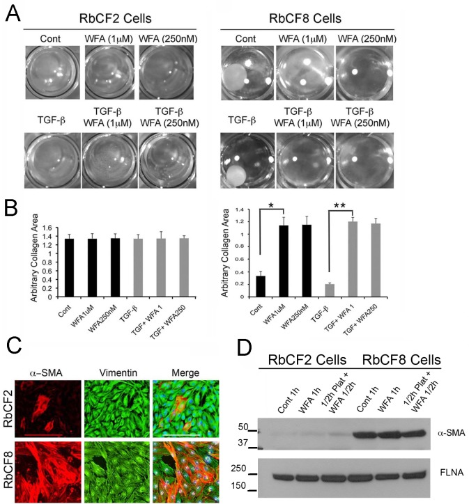
Fig 8. WFA controls gel contraction properties in a cell-passage dependent manner.
(A) Representative images of RbCF2 and RbCF8 cells embedded in collagen type I gels and treated with TGF-β1 (2 ng/ml) for 2 days in presence or absence of different doses of WFA. Gels were then fixed and images were captured at 2X magnification. (B) Quantification of gel contractile activity. Images from (A) were used to arbitrarily measure the collagen area in each well using ImageJ program (n = 8 wells/treatment). (C) Immunohistochemistry of RbCF2 and RbCF8 cells cultured on slides and stained with α-SMA (red), anti-vimentin (green) and DAPI (blue). Scale bar = 215 μm at 10X magnification. (D) Western blot analysis of RbCF2 and RbCF8 cells for α-SMA expression. Cells were trypsinized and plated for 30 min and then treated with 1 μM WFA for 30 min or treated with WFA at time of plating for 1 h. Blots were probed with anti-α-SMA and FLNA antibodies used as loading control.

