
Fig 10. WFA blocks activation of the ERK1/2 pathway in vivo by targeting pSer38vim expression.
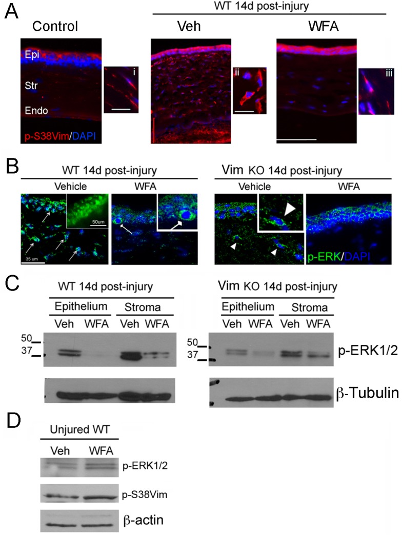
Anesthetized mice were subjected to bilateral corneal alkali injury. Injured mice were treated with DMSO (Vehicle) or WFA (2 mg/kg solubilized in DMSO) on the day of injury and every subsequent day by intraperitoneal injection for 14 days. (A) Immunohistochemistry analysis of WT corneal sections stained with pSer38vim antibody (red) and DAPI (blue). Epi: Epithelium; Str: Stroma; End: Endothelium. Enlarged panels: 60X magnification. (B) Immunohistochemistry analysis of WT and Vim KO samples stained with anti-pERK1/2 antibody (Green) and DAPI (blue). Enlarged panel: 60X magnification of WT-Vehicle sample, showing nuclear expression pERK1/2 (Green). Pointed-ended arrows indicate nuclear localization of pERK1/2 in epithelial and stromal cells of WT-Vehicle samples. Square-ended arrow indicates cytoplasmic localization of pERK1/2 in the basal layer of the epithelium of WT-WFA samples. Arrowhead indicates partial cytoplasmic localization of pERK1/2 in stromal cells of Vim KO-Vehicle samples. (C) Western blot analysis of epithelial and stromal extracts from corneas of mice 14 d post-injury. Equal amount of total protein extracts were fractionated by SDS-PAGE and blots were probed with anti-pERK1/2 antibody. β-tubulin was used as loading control. (D) Western blot analysis of corneal extracts from uninjured WT mice treated for 7 d with WFA (2 mg/kg/d) or vehicle (DMSO). Total protein extracts were fractionated by SDS-PAGE and blots were probed with anti-vimentin, and anti-pERK1/2 antibodies. β-actin antibody was used as loading control.