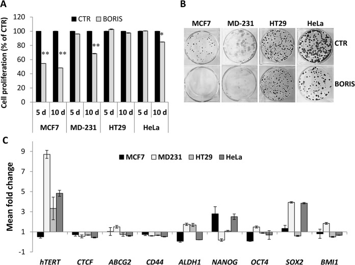
Fig 9. Inhibition of cell growth and up-regulation of hTERT and stem cell genes expression, after BORIS-induction in epithelial cancer cells.
MCF7, MDA-MB-231, HT29 and HeLa cells were engineered to stably express BORIS cDNA. After transduction with either lentivirus harboring BORIS cDNA (BORIS) or control lentivirus (CTR), cells were selected by incubation with the antibiotic G418 for at least 2 weeks. (A) Cell proliferation was analyzed by MTT assay after 5 and 10 days of dox-induction of BORIS expression. Results are indicated as a percentage compared to the control cells (CTR). Error bars represent the mean ± SD (n = 3). One asterisk (p<0.05) or two asterisks (p<0.01) indicate statistically significant difference between BORIS and CTR cells. (B) Representative images of colony formation assay. Three hundred cells were seeded in each well of 6-well plates with medium containing doxycycline, each assay was performed in triplicate. Cells were cultured for 2 weeks, then were fixed and stained with crystal violet. (C) After 2 weeks of dox-induction of BORIS and CTR cells, mRNA levels of the indicated genes were analyzed by qRT-PCR. Graph represents for each gene the fold induction of BORIS-induced cells related to control cells. Error bars represent the mean ± SD (n = 3).

