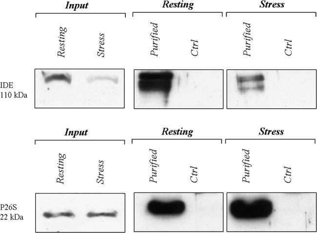
Fig 7. Decreased IDE-proteasome interaction during the HSR.

A semi-quantitative comparison of IDE association with the proteasome by Western blotting. Membranes were stained with an anti-IDE antibody (upper panel) and with an anti-protesaome (p27 subunit) antibody (lower panel). Input, stands for 5 micrograms of cytosolic extract loaded in the gel, whereas Ctrl stands for incubation of cell lysates with non-specific beads. A representative immunoblot of three independent experiment is shown. Proteasome particles from the cytosolic fraction of resting and heat-stressed SHSY5Y were affinity purified. Notably, a lower molecular weight species is recognized by the anti-IDE antiboby. Anyway, we are unsure about the identity of this band which, anyway, is constantly observed by us and by other authors when large amount of cell extracts is loaded.
