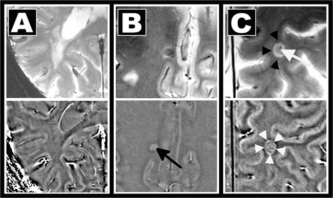
Fig 1. Methods.

MS lesion morphology was assessed on T2*w (top) and phase (bottom) images using a visual analysis. Lesion visibility, the existence of a central vein, and signal alterations indicative for iron deposition were rated. A: confluent T2*w hyperintense lesion not visible on phase images. B: central T2*w hypointensity and positive phase shift indicative of iron deposits (arrow). C: Iron deposits in a perivascular (white arrow) lesion causing a T2*w hypointense rim (black arrowheads) and ring-like paramagnetic (“bright”) phase alterations (white arrowheads). Please note that the dark envelop around the “bright” ring in the phase image likely represents an artifact caused by the dipolar field patterns.
