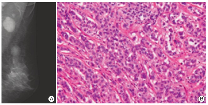
Fig. 1.

Radiologic and histologic features of the tumors. (A) The medial lateral oblique mammogram of the left breast shows 2×1.7-cm-sized oval shaped isodense mass with partially obscured margin at upper outer quadrant and 3.4×2.3-cm-sized enlarged axillary lymph node with loss of radiolucent fatty hilum at left axilla. (B) Invasive ductal carcinoma of breast (nuclear grade 2, histologic grade 2) reveals irregular infiltrative nests of tumor cells (H&E staining, ×200).
