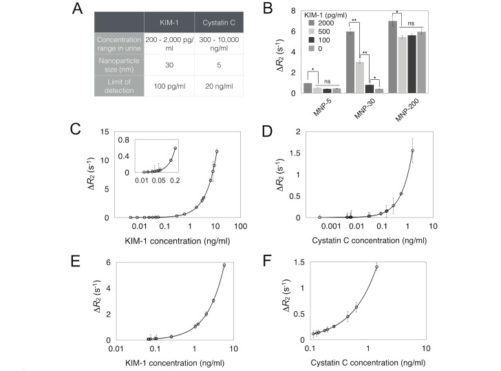
Fig 2. Assay validation.
A, Assay set-up for urinary marker detection. B, μNMR signals using MNPs with different sizes. * = p < 0.02, ** = p < 0.001, ns = not significant. Detection sensitivity measurements using serial dilutions of recombinant KIM-1(C) and Cystatin C (D) in buffer solution. Inset in C shows data points in low ranges of KIM-1. ΔR2 = R2 (sandwich)—R2 (bead only). E,F, Detection of KIM-1 (E) and Cystatin C (F) in 100% and 20% urine, respectively. The urine used was from a healthy patient (no. 20) with ranges of KIM-1 and Cystatin C that were not detectable. Note that all the clinical samples (in Fig 3) were analyzed based on the same dilution factors.

