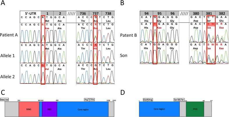
Fig 2. RAG1/2 Mutation analysis of genomic DNA from peripheral blood.
(A) Patient A is compound heterozygous in RAG1, c.1 A>G, p.M1V; c.2322 G>A, p.R737H. Allele-specific PCR was used to characterize RAG1 M1V and R737H alleles. (B) Patient B is compound heterozygous in RAG2, c1347-8delCT, pSer381Terfs*1; c488G>A, pGlu95R. Mutation analysis of the son of patient B identified the compound heterozygosity of the RAG2 missense mutations. (C) Schematic depiction of RAG1 (D) Schematic depiction of RAG2. Mutations are indicated by rectangles.

